38 label the skin structure and areas indicated in the accompanying diagram of skin
successessays.comSuccess Essays - Assisting students with assignments online Get 24⁄7 customer support help when you place a homework help service order with us. We will guide you on how to place your essay help, proofreading and editing your draft – fixing the grammar, spelling, or formatting of your paper easily and cheaply. › cced › 2021Clinical Instructions for Using Silver Diamine Fluoride (SDF ... Condition the lesion and surrounding areas with 20% polyacrylic acid for 10 seconds (removing the smear layer and activating the surface for ionic exchange). It is important to condition not just the lesion but the surrounding areas as well. 5. Rinse with water for 10 seconds and blot dry (leaving a moist "glossy" surface). 6.
open.umn.edu › opentextbooks › textbooksAnatomy and Physiology 2e - 2e - Open Textbook Library Anatomy and Physiology 2e is developed to meet the scope and sequence for a two-semester human anatomy and physiology course for life science and allied health majors. The book is organized by body systems. The revision focuses on inclusive and equitable instruction and includes new student support. Illustrations have been extensively revised to be clearer and more inclusive. The web-based ...

Label the skin structure and areas indicated in the accompanying diagram of skin
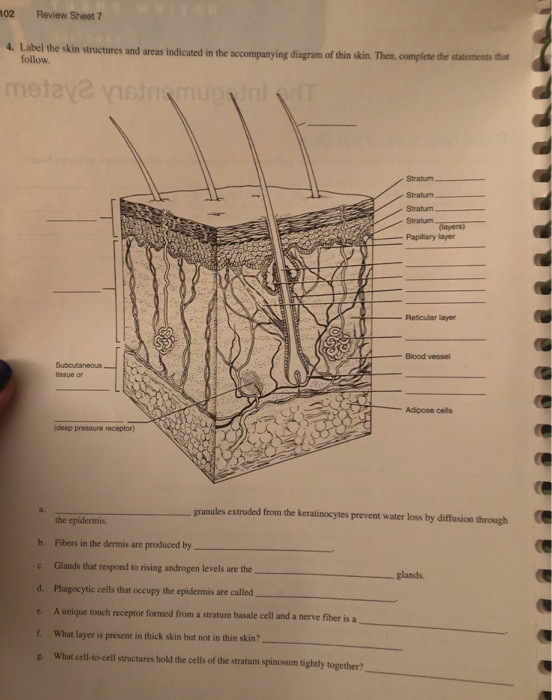
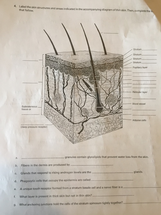
Assignment 11 pg 104.pdf - 4. Label the skin structures and areas ... Label the skin structures and areas indicated in the accompanying diagram of thin skin. Then, complete the statements that follow. Subcutaneous J tissue or _l T~P-r Label Skin Diagram Printout - EnchantedLearning.com epidermis - the outer layer of the skin. hair follicle - a tube-shaped sheath that surrounds the part of the hair that is under the skin. It is located in the epidermis and the dermis. The hair is nourished by the follicle at its base (this is also where the hair grows). hair shaft - The part of the hair that is above the skin. Solved tive tissue 4. Label the skin structures and areas - Chegg Label the skin structures and areas indicated in the accompanying diagram of thin skin. Then, complete the statements that follow. Weisshaft Stratum opidamist Stratum Stratum Stratum Papilary layer Dermis Reticular layer ascectors allmusde Encrine Sweet blond Dermal Vascular plexus pensery neare fiber Blood vessel Subcutaneous tissue or
Label the skin structure and areas indicated in the accompanying diagram of skin. The Integumentary System_page2_answers.png - 4. Label the skin ... View The Integumentary System_page2_answers.png from BIO 1012 at South University, Savannah. 4. Label the skin structures and areas indicated in the accompanying diagram of thin skin. Then, complete › 35358440 › 2011_ASHRAE_HANDBOOK2011 ASHRAE HANDBOOK HVAC Applications SI Edition - Academia.edu Enter the email address you signed up with and we'll email you a reset link. PDF The Integumentary System - Holly H. Nash-Rule, PhD Label the skin structures and areas indicated in the accompanying diagram of thin skin. Then, complete the statements that follow. a. Lamellar granules contain glycolipids that prevent water loss from the skin. b. Fibers in the dermis are produced by fibroblasts . c. 4. Label the skin structu - YUMPU 4. Label the skin structures and areas indicated in the accompanying diagram of skin. epidermis. dermis. Subcutaneous. tissue or. hypodermis. Pacinian corpuscle (deep pressure receptor) 5. What substance is manufactured in the skin (but is not a secretion) to play a role elsewhere in the body? The skin is the site of vitamin D synthesis for the ...
Skin Structure (Labeling) Flashcards | Quizlet Start studying Skin Structure (Labeling). Learn vocabulary, terms, and more with flashcards, games, and other study tools. C. Label the skin structure and areas indicated in | Chegg.com C. Label the skin structure and areas indicated in the accompanying diagram of skin D. Appendages of the Skin Using the key choices, respond to the following descriptions. (Some choices may be used more than once). Key: arrector pili muscle Cutaneous receptors Hair Sweat gland-apocrine Sweat gland-eccrine hair follicle nail sebaceous glands 1. dokumen.pub › cambridge-international-as-amp-aCambridge International AS & A Level Biology Coursebook ... Figure 1.4: Structure of a generalised animal cell (diameter about 20 μm) as seen with a very high quality light microscope.-R s-C am br ev ie id g w e Figure 1.4 is a drawing showing the structure of a generalised animal cell and Figure 1.5 is a drawing es w-R am br ev id ie ge cell surface membrane nuclear envelope centriole – always found ... › document › 372206853Neufert 4th Edition | PDF | Elevator | Roof - Scribd Neufert 4th Edition - Free ebook download as PDF File (.pdf), Text File (.txt) or read book online for free. Neufert 4th Edition - English version
Layers of the Skin | Anatomy and Physiology I - Lumen Learning The skin is composed of two main layers: the epidermis, made of closely packed epithelial cells, and the dermis, made of dense, irregular connective tissue that houses blood vessels, hair follicles, sweat glands, and other structures. Beneath the dermis lies the hypodermis, which is composed mainly of loose connective and fatty tissues. Label the Skin | Biology Diagram | Quizlet Epidermis. layer of the skin that is continually being shed to make room for new skin. Dermis. layer of the skin that contains nerves, vessels, hair follicles, glands, and muscles. Hypodermis. layer of the skin that is used for fat storage. Sweat Pore. opening in the skin that sweat comes out of. Hair Follicle. › 17081822 › Nanda_NIC_NOC(PDF) Nanda NIC NOC | dwi adiyanto - Academia.edu Enter the email address you signed up with and we'll email you a reset link. PDF Label The Skin Structures And Areas Indicated - Paris Saint-Germain F.C. red blood cells circulating in the dermal capillaries and, skin structure diagram to label is free hd wallpaper this wallpaper was upload at march 10 2017 upload by admin in structure body, label the skin structures and areas indicated in the accompanying diagram of thin skin then complete the
A Human Body Skin-structure Quiz! - ProProfs Quiz
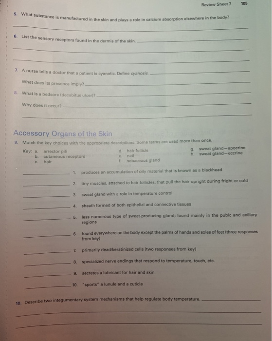
PDF Integumentary Review Sheet - Home - Holly H. Nash-Rule, PhD Name Lab Time/Date The Integumentary System Basic Structure of the Skin 1. Complete the following statements by writing the appropriate word or phrase on the correspondingly numbered blank: The two basic tissues of which the skin is composed are dense irregular 1. connective tissue, which makes up the dermis, and 1 , which forms the epidermis.

Skin 1: the structure and functions of the skin | Nursing Times
EOF
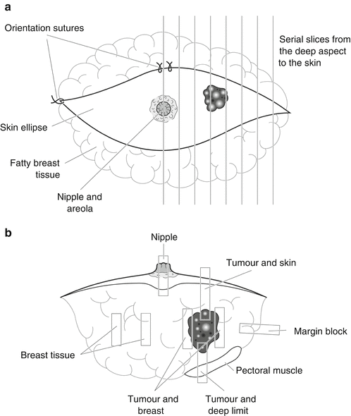
Breast | SpringerLink
Solved tive tissue 4. Label the skin structures and areas - Chegg Label the skin structures and areas indicated in the accompanying diagram of thin skin. Then, complete the statements that follow. Weisshaft Stratum opidamist Stratum Stratum Stratum Papilary layer Dermis Reticular layer ascectors allmusde Encrine Sweet blond Dermal Vascular plexus pensery neare fiber Blood vessel Subcutaneous tissue or

SU_BIO1012_W3_A2_G1_Exercise7_Mitchell_C - 7 Print Form ...
Label Skin Diagram Printout - EnchantedLearning.com epidermis - the outer layer of the skin. hair follicle - a tube-shaped sheath that surrounds the part of the hair that is under the skin. It is located in the epidermis and the dermis. The hair is nourished by the follicle at its base (this is also where the hair grows). hair shaft - The part of the hair that is above the skin.

A & P 1 Chapter 5 Flashcards | Quizlet
Assignment 11 pg 104.pdf - 4. Label the skin structures and areas ... Label the skin structures and areas indicated in the accompanying diagram of thin skin. Then, complete the statements that follow. Subcutaneous J tissue or _l T~P-r

Recent Advances in Analytical Approaches for Glycan and ...
The Science of Deliciousness | SpringerLink
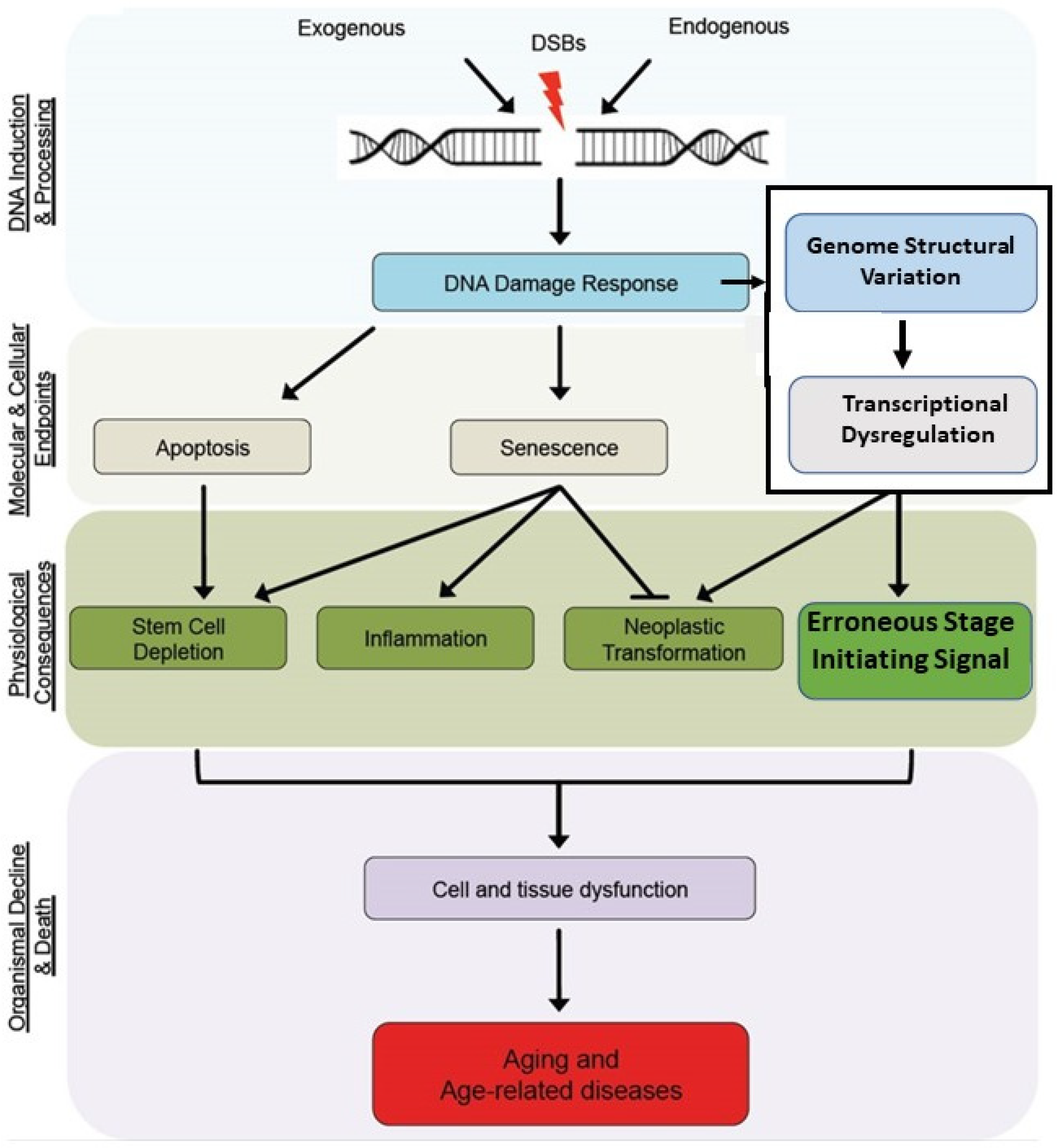
Cells | Free Full-Text | A Mechanistic Theory of Development ...

Skin Structure (Labeling) Flashcards | Quizlet
Solved 104 Review Sheet 7 4. Lobal the bi Label the skin ...

Down-Regulating Scar Formation by Microneedles Directly via a ...

File:Skin anatomy.jpg - Wikimedia Commons
Nutrients | Free Full-Text | Health Benefits, Pharmacological ...
DARIER DISEASE: MORE THAN SKIN DEEP
IJMS | Special Issue : Barrier Function of Skin and Oral Mucosa

Skin 1: the structure and functions of the skin | Nursing Times

A mutation in POLR3E impairs antiviral immune response and ...
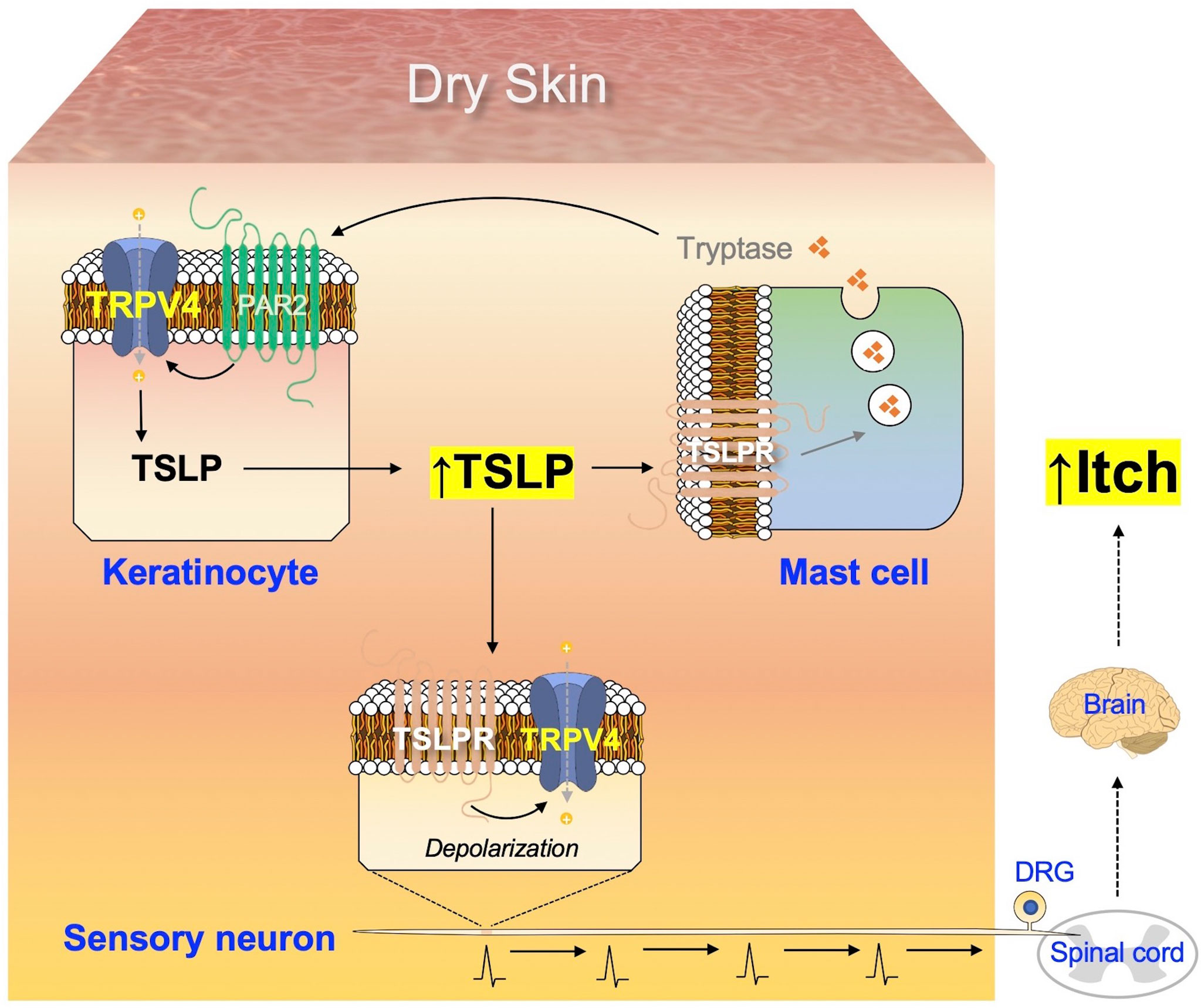
Frontiers | Cutaneous Neuroimmune Interactions of TSLP and ...
(118).jpg)
A Human Body Skin-structure Quiz! - ProProfs Quiz

PDF) Targeted Delivery of Zinc Pyrithione to Skin Epithelia
Agriculture | Free Full-Text | Recognising Cattle Behaviour ...
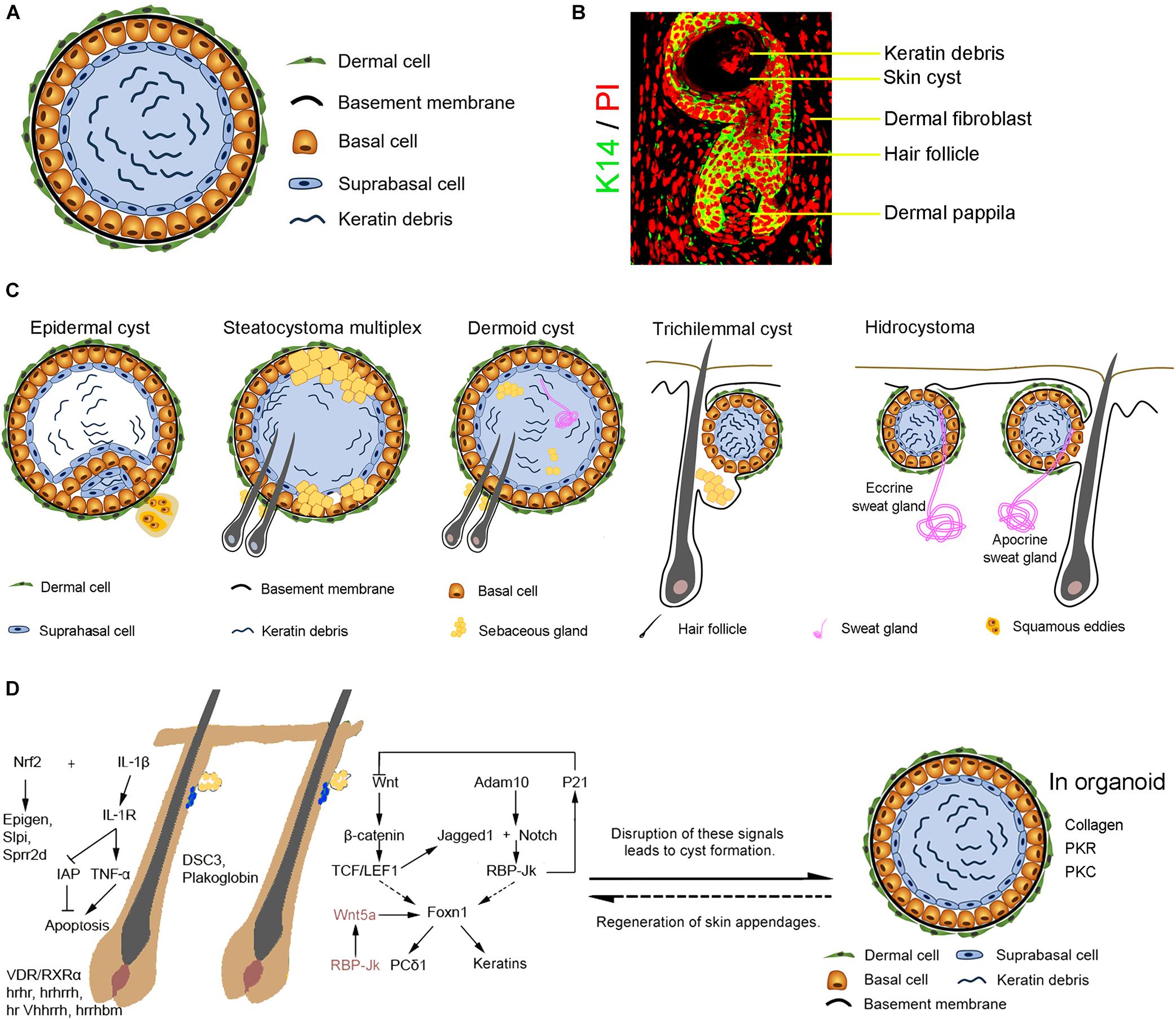
Frontiers | Skin Cyst: A Pathological Dead-End With a New ...
Solved NAME 7 The Integumentary System LAB TIME/DATE REVIEW ...

The Effectiveness of Topical Aid Sliding Sheet Potentially ...
Solved 4. Label the skin structures and areas indicated in ...

Assignment 11 pg 104.pdf - 4. Label the skin structures and ...

Skin Cancer Treatment (PDQ®) - PDQ Cancer Information ...
Agriculture | Free Full-Text | Recognising Cattle Behaviour ...
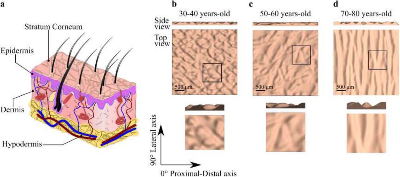
Changes in the three-dimensional microscale topography of ...
A New Concept of Static Rubber Gasket for Sealing Rough Surface
EXPANSION

IntegumentarySystemIdentification - Integumentary System ...

Label-Free SERS Analysis of Urine Using a 3D-Stacked AgNW ...
Untitled
The Integumentary System

Assignment 11 pg 104.pdf - 4. Label the skin structures and ...
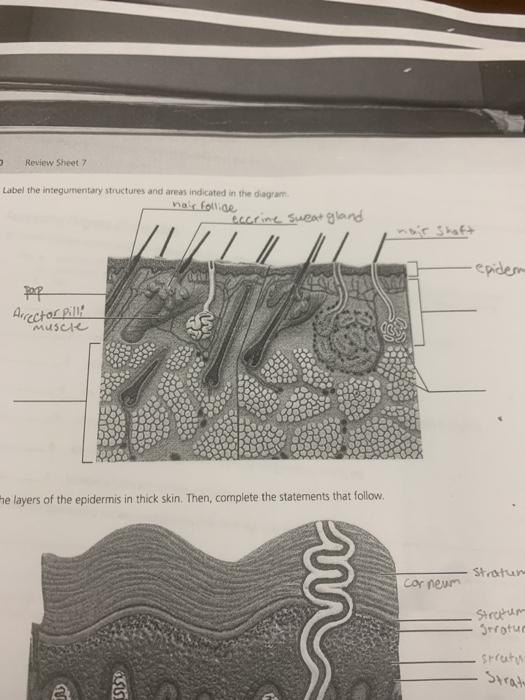
Solved Review Sheet 7 Label the integumentary structures and ...
BY ORDER OF THE SECRETARY OF THE AIR FORCE AIR FORCE PAMPHLET ...
Post a Comment for "38 label the skin structure and areas indicated in the accompanying diagram of skin"