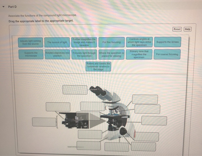
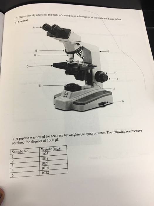
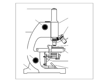
42 drag the labels onto the diagram to identify the parts of the compound microscope (1 of 2).
Labels The The To Structures. Drag Onto The Diagram Identify Different Types of DNA in Different Organisms 8 Petharbor Redlands From the capsule, strands of tissue pass into Part A Drag the labels onto the diagram to identify the directional terms (2 of 2) Part A Drag the labels onto the diagram to identify the directional terms (2 of 2). Match the rational numbers with their equivalent forms Drag the ... › en › productHow to run an assay | Agilent Important: For Induced XF Glycolytic Rate Assay (1 or 2 injections prior to standard injections of rontenone/antimycin A and 2-DG),, you must identify the Rotenone/Antimycin-A injection using the drop-down menu seen above the widget before you can add this analysis view. This Rotenone/Antimycin-A injection selection plays a critical role in ...
quizlet.com › 524370406 › a-p-microscope-and-cellA & P Microscope and Cell cycle Flashcards | Quizlet Study with Quizlet and memorize flashcards containing terms like What is the typical magnification provided by the eyepiece (ocular) lens system?, The greatest magnification on the compound light microscope can be achieved by using the high power objective lens., A parfocal microscope is one that keeps the specimen in focus (or very close to it) when a higher-power objective lens is rotated ...

Drag the labels onto the diagram to identify the parts of the compound microscope (1 of 2).
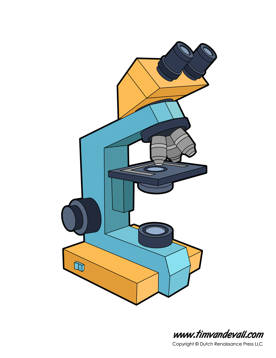
Compound Microscope - Types, Parts, Diagram, Functions and Uses It comes with a wide body and base. Its distinct parts include a condenser, illumination, focus lock, mechanical stage, and a revolving nosepiece which can hold up to five objectives. It usually has a binocular head, which makes long-term observation easy. Image 22: An example of a research compound microscope. Microscope Parts and Functions Body tube (Head): The body tube connects the eyepiece to the objective lenses. Arm: The arm connects the body tube to the base of the microscope. Coarse adjustment: Brings the specimen into general focus. Fine adjustment: Fine tunes the focus and increases the detail of the specimen. Nosepiece: A rotating turret that houses the objective lenses. Compound Microscope: Parts of Compound Microscope - BYJUS (A) Mechanical Parts of a Compound Microscope 1. Foot or base It is a U-shaped structure and supports the entire weight of the compound microscope. 2. Pillar It is a vertical projection. This stands by resting on the base and supports the stage. 3. Arm The entire microscope is handled by a strong and curved structure known as the arm. 4. Stage
Drag the labels onto the diagram to identify the parts of the compound microscope (1 of 2).. Compound Microscope Parts - Labeled Diagram and their Functions The eyepiece (or ocular lens) is the lens part at the top of a microscope that the viewer looks through. The standard eyepiece has a magnification of 10x. You may exchange with an optional eyepiece ranging from 5x - 30x. [In this figure] The structure inside an eyepiece. The current design of the eyepiece is no longer a single convex lens. Parts of a microscope with functions and labeled diagram - Microbe Notes Q. List down the 18 parts of a Microscope. 1. Ocular Lens (Eye Piece) 2. Diopter Adjustment 3. Head 4. Nose Piece 5. Objective Lens 6. Arm (Carrying Handle) 7. Mechanical Stage 8. Stage Clip 9. Aperture 10. Diaphragm 11. Condenser 12. Coarse Adjustment 13. Fine Adjustment 14. Illuminator (Light Source) 15. Stage Controls 16. Base 17. Binocular Microscope Anatomy - Parts and Functions with a Labeled Diagram Non-optical parts of the compound microscope. The important non-optical parts of the light compound microscope are the body tube or head, arm or frame, fine adjustment, coarse adjustment, nose piece, stage, and base. Now, I will describe all these non-optical parts of the light compound microscope with the labeled diagrams. Parts of the Microscope with Labeling (also Free Printouts) Let us take a look at the different parts of microscopes and their respective functions. 1. Eyepiece it is the topmost part of the microscope. Through the eyepiece, you can visualize the object being studied. Its magnification capacity ranges between 10 and 15 times. 2. Body tube/Head It is the structure that connects the eyepiece to the lenses.
Bio2514 Week 3 The Microscope - Lab Topic.docx - Course Hero Drag the labels onto the diagram to identify the parts of the compound microscope (1 of 2). Arm ocular lens Mechanical stage rotating nose piece Stage Objective lenses Condenser Iris diaphragm lever 3. The microscope slide rests on the __________ while being viewed. Stage 4. Your lab microscope is parfocal. label parts of a compound microscope - TeachersPayTeachers Students will label the parts of the microscope, calculate total magnification, determine measurements, answer questions about the proper use of the microscope, and much more.Choose to use the traditional printable version, or the paperless, digital Google Apps version. This resource is perfect for traditional classrooms, distance learning, and. Mastering Biology Chp. 8 HW - Quizzes Studymoose PART B - Energetics of electron transport This diagram shows the basic pattern of electron transport through the four major protein complexes in the thylakoid membrane of a chloroplast. ELECTRON TRANSPORT STEP: 1. Water → P680⁺ 2. P680 → Pq (plastoquinone) 3. Pq → P700⁺ 4. P700 → Fd (ferredoxin) 5. Fd → NADP⁺ Labeling the Parts of the Microscope | Microscope activity, Science ... Print a microscope diagram, microscope worksheet, or practice microscope quiz in order to learn all the parts of a microscope. Tim's Printables. Science Printables. ... Jan 13, 2016 - Free worksheets for labeling parts of the microscope including a worksheet that is blank and one with answers. Jan 13, 2016 - Free worksheets for labeling parts ...
› de › jobsFind Jobs in Germany: Job Search - Expatica Germany Browse our listings to find jobs in Germany for expats, including jobs for English speakers or those in your native language. 16 Parts of a Compound Microscope: Diagrams and Video Base of the Microscope 4. Eyepiece The eyepiece, also known as the "Ocular", is the first magnification lens you will look through in a compound microscope. Put simply, this is where you put your eye to see the image. Usually eyepieces come in 10X magnification, or 15X magnification but they can vary from 5X - 30X. Compound Microscope Labeled Diagram | Quizlet Part that supports the microscope. Stage Supports the slide or specimen Coarse adjustment Knob sed to focus when using the low power objective lenses Fine Adjustment Knob Used to focus the image on high power to view image in more detail. Revolving nose piece The revolving piece on which the lenses are attached quizlet.com › 512343334 › ap-lab-3-hw-flash-cardsa&p lab 3 hw Flashcards | Quizlet Before putting away the microscope in the storage cabinet you must observe all of the following except ________. rotate the highest power objective lens in position. Drag the labels onto the diagram to identify the parts of the compound microscope (1 of 2). (figure 3.1) left column: arm. mechanical stage.
Answer correct score summary your score on this - Course Hero Drag the labels to identify the parts of the compound microscope. Not all labels will be used. Hint 1.Objective and ocular lenses Both objective and ocular lenses magnify the specimen. Objective lenses are nearest the specimen and can be rotated to increase magnification. Ocular lenses are nearestthe eye and have a fixed magnification.
Compound Microscope: Definition, Diagram, Parts, Uses, Working ... - BYJUS The parts of a compound microscope can be classified into two: Non-optical parts Optical parts Non-optical parts Base The base is also known as the foot which is either U or horseshoe-shaped. It is a metallic structure that supports the entire microscope. Pillar The connection between the base and the arm are possible through the pillar. Arm
Labeling the Parts of the Microscope | Microscope World Resources Labeling the Parts of the Microscope This activity has been designed for use in homes and schools. Each microscope layout (both blank and the version with answers) are available as PDF downloads. You can view a more in-depth review of each part of the microscope here. Download the Label the Parts of the Microscope PDF printable version here.
Compound Microscope: Parts of Compound Microscope - BYJUS (A) Mechanical Parts of a Compound Microscope 1. Foot or base It is a U-shaped structure and supports the entire weight of the compound microscope. 2. Pillar It is a vertical projection. This stands by resting on the base and supports the stage. 3. Arm The entire microscope is handled by a strong and curved structure known as the arm. 4. Stage
Microscope Parts and Functions Body tube (Head): The body tube connects the eyepiece to the objective lenses. Arm: The arm connects the body tube to the base of the microscope. Coarse adjustment: Brings the specimen into general focus. Fine adjustment: Fine tunes the focus and increases the detail of the specimen. Nosepiece: A rotating turret that houses the objective lenses.
Compound Microscope - Types, Parts, Diagram, Functions and Uses It comes with a wide body and base. Its distinct parts include a condenser, illumination, focus lock, mechanical stage, and a revolving nosepiece which can hold up to five objectives. It usually has a binocular head, which makes long-term observation easy. Image 22: An example of a research compound microscope.






(22).jpg)








Post a Comment for "42 drag the labels onto the diagram to identify the parts of the compound microscope (1 of 2)."